
Imaging of the human auditory cortex, to date, has been somewhat restricted by the limitations of traditional neuroimaging methodologies, such as the noisiness of magnetic resonance imaging (MRI) that often interferes with the presentation of experimental sounds.

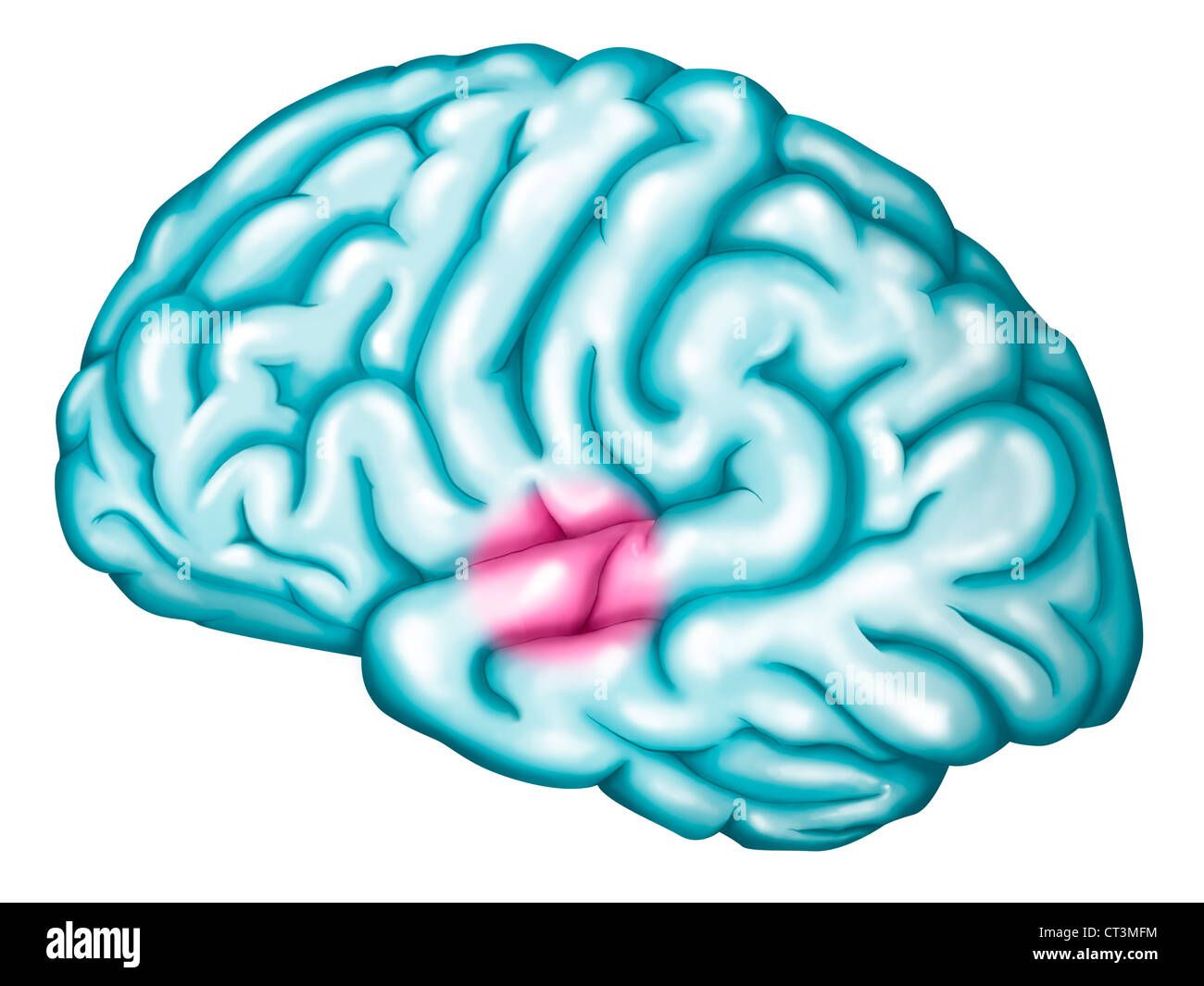
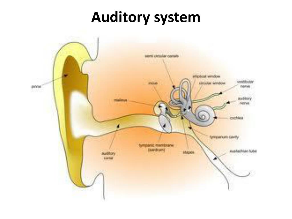
As its name suggests, the auditory cortex’s primary role is to process incoming auditory signals – this can include speech, non-speech sounds and music. Together these areas form the auditory cortex of the human brain. Surrounding it is the auditory association area. It is situated in the superior temporal gyrus (STG) and extends into Heschl’s gyrus and the lateral sulcus. The primary auditory cortex is located bilaterally in the temporal lobes, and corresponds to Brodmann areas 41 and 42. Keywords: optical imaging, hearing loss, superior temporal gyrus, plasticity, auditory processing In particular, the benefits and limitations of using functional near infrared spectroscopy (fNIRS) on complex populations such as infants and individuals with hearing loss are explored, along with suggestions for future research developments. This review presents a brief history of optical imaging, followed by an exploration of how advances in optical imaging technologies have increased the understanding of the functions and processes within the auditory cortex. Optical imaging provides a new and exciting option for exploring this key cortical area. NIHR Biomedical Research Centre, Ropewalk House, 113 The Ropewalk, Nottingham NG1 5DU, UKĪbstract: Imaging the auditory cortex can prove challenging using neuroimaging methodologies due to interfering noise from the scanner in fMRI and the low spatial resolution of EEG. Samantha C Harrison, 1, 2 Douglas EH Hartley 1–3ġNIHR Nottingham Biomedical Research Centre, Nottingham, UK 2Hearing Sciences Group, Division of Clinical Neuroscience, School of Medicine, University of Nottingham, Nottingham, UK 3Department of Otolaryngology, Nottingham University Hospitals National Health Service Trust, Nottingham, UK


 0 kommentar(er)
